Fichye:DTI-sagittal-fibers.jpg
Dimansyon previzyalizasyon sa a : 643 × 600 piksèl. Lòt rezolisyon yo: 257 × 240 piksèl | 515 × 480 piksèl | 1 021 × 952 piksèl.
Fichye orijinal (1 021 × 952 piksèl, gwosè fichye a : 294 kio, tip MIME li ye : image/jpeg)
|
Rezime
| Deskripsyon |
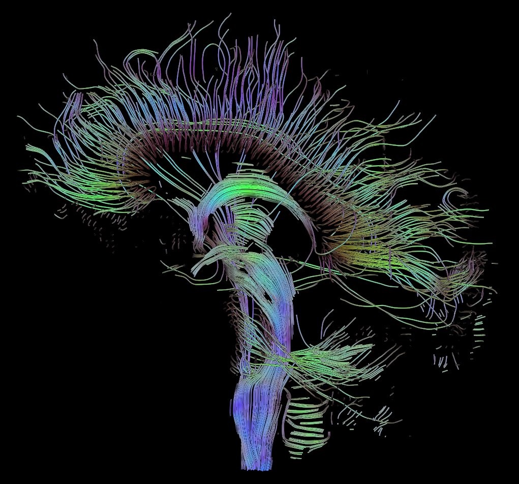
English: Visualization of a DTI measurement of a human brain. Depicted are reconstructed fiber tracts that run through the mid-sagittal plane. Especially prominent are the U-shaped fibers that connect the two hemispheres through the corpus callosum (the fibers come out of the image plane and consequently bend towards the top) and the fiber tracts that descend toward the spine (blue, within the image plane)
Français : Visualisation d'une mesure DTI d'un cerveau humain. Ce qui est représenté sont des faisceaux de fibres reconstruits qui traversent le plan demi-sagittal. On observe les fibres en U qui connectent les deux hémisphères à travers le corps calleux, qui sont particulièrement importantes (les fibres sortent du plan de l'image et par conséquent se courber vers le haut) ainsi que les faisceaux de fibres qui descendent vers la colonne vertébrale (bleu, dans le plan de l'image)
Deutsch: Traktographie-Verfahren rekonstruieren aus den Messdaten der Diffusions-Tensor-Bildgebung den anzunehmenden Verlauf größerer Nervenbahnen. Hier dargestellt sind die Ergebnisse für ein menschliches Gehirn; um die Übersichtlichkeit zu wahren, beschränkt sich die Abbildung auf Bahnen, die die Medianebene schneiden. Insbesondere sind dies die U-förmigen Faserbündel, die die beiden Hirnhälften verbinden (sie durchstoßen die Bildebene und sind nach oben gebogen) sowie die Faserbündel, die zum Rückenmark ziehen (blau dargestellt, liegen innerhalb der Bildebene) |
| Dat | |
| Sous | Travay pèsonèl |
| Kreyatè | Thomas Schultz |
| Pèmisyon : (Resèvi ak fichye sa a) |
Rendering is own work, using a modified version of the BioTensor application developed at the University of Utah. The dataset is courtesy of Gordon Kindlmann at the Scientific Computing and Imaging Institute, University of Utah, and Andrew Alexander, W.M. Keck Laboratory for Functional Brain Imaging and Behaviour, University of Wisconsin, Madison. It is publicly available from [1] |
Kondisyon itlizasyon
Moi, en tant que détenteur des droits d’auteur sur cette œuvre, je la publie sous les licences suivantes :

|
Ou bay pèmisyon pou kopye, distribye ak/oswa modifye dokiman sa a dapre kondisyon GNU Free Documentation License, Vèsyon. 1.2 oswa nenpòt vèsyon ki pibliye pita pa Free Software Foundation; san okenn Seksyon ki pa ka chanje, pa gen okenn Tèks ak premye paj kouvèti, epi pa gen okenn Tèks Denyè-Kouvèti. Gen yon kopi lisans lan nan seksyon ki rele GNU Free Documentation License. |
| Fichye sa a distribiye selon lisans Creative Commons. | ||
| ||
| Ce bandeau de licence a été ajouté à ce fichier dans le cadre de la procédure de mise à jour des licences des images sous GFDL. |
Ce fichier est sous licence Creative Commons Attribution – Partage dans les Mêmes Conditions 2.5 Générique, 2.0 Générique et 1.0 Générique.
- Ou gen dwa:
- pou pataje – pou kopye, distribiye epi difize ouvraj la
- pou remiks – pou adapte ouvraj la
- Selon kondisyon sa a yo:
- Rekonèt avèk non otè – Ou dwe bay enfòmasyon apwopriye sou otè a, bay yon lyen pou lisans lan, epi endike si te gen chanjman ki fèt. Ou ka fè sa nan nenpòt fason rezonab, men okenn fason ki sijere ke otè a soutni oswa apwouve itilizasyon ou an.
- Enfòmasyon sa yo ka pataje – Si ou fè rekopye, transfòme, oswa baze sou materyèl sa, ou dwe distribye kontribisyon ou anba menm oswa yon lisans konpatib avèk orijinal la.
Chwazi lisans ou vle a.
Istorik fichye a
Klike sou yon dat/yon lè pou wè fichye a jan li te ye nan moman sa a.
| Dat ak lè | Minyati | Grandè yo | Itilizatè | Komantè | |
|---|---|---|---|---|---|
| Kounye a | 13 oktòb 2017 à 10:42 | | 1 021 × 952 (294 kio) | Mikael Häggström | Minor crop of black areas at the top and bottom |
| 22 septanm 2006 à 16:22 |  | 1 021 × 1 125 (203 kio) | Thomas Schultz | {{Information |Description=Visualization of a DTI measurement of a human brain. Depicted are reconstructed fiber tracts that run through the mid-sagittal plane. Especially prominent are the U-shaped fibers that connect the two hemispheres through the corp |
Itilizasyon fichye sa a
paj sa a itilize fichye sa a:
Itilizasyon global fichye a
Wiki sa a yo sèvi ak fichye sa a:
- Itilizasyon sou af.wikipedia.org
- Itilizasyon sou ar.wikipedia.org
- Itilizasyon sou az.wikiquote.org
- Itilizasyon sou bn.wikipedia.org
- Itilizasyon sou cs.wikipedia.org
- Itilizasyon sou de.wikipedia.org
- Autismus
- Computergrafik
- Bipolare Störung
- Portal:Informatik/Exzellente Artikel
- Portal:Geist und Gehirn/Artikel des Monats
- Diffusions-Tensor-Bildgebung
- Wikipedia:Kandidaten für exzellente Bilder/Archiv2006/17
- Datei:DTI-sagittal-fibers.jpg
- Wikipedia:Exzellente Bilder/Naturwissenschaften
- Portal:Physik/Artikel des Monats 2024-03
- Wikipedia:Exzellente Bilder/Kleine Bilder
- Itilizasyon sou en.wikipedia.org
- Neurolinguistics
- Tractography
- Portal:Medicine
- User talk:Spikebrennan
- User:Spikebrennan
- Diffusion MRI
- Wikipedia:WikiProject Neuroscience
- Portal:Psychology/Selected article
- Wikipedia:Featured pictures/Sciences/Biology
- Portal:Psychology/Selected article/7
- Wikipedia:Featured pictures thumbs/08
- Wikipedia:Featured picture candidates/DTI-sagittal-fibers.jpg
- Wikipedia:Wikipedia Signpost/2007-11-05/Features and admins
- Wikipedia:Featured picture candidates/November-2007
- Wikipedia:Picture of the day/March 2008
- Connectome
- Template:POTD/2008-03-10
- User talk:Thomas Schultz
- Wikipedia:Wikipedia Signpost/2007-11-05/SPV
- Biological data visualization
- Wikipedia:WikiProject Medicine/Recognized content
- Wikipedia:WikiProject Molecular Biology/Biophysics
- User:Wouterstomp/test
- Wikipedia:WikiProject Anatomy/Resources
- Wikipedia:WikiProject Anatomy/Recognized content
- Wikipedia talk:WikiProject Anatomy/Archive 9
- Portal:Medicine/Recognized content
- User talk:Rhododendrites/Reconsidering FPC on the English Wikipedia
- User:Hydrogenkitsch
- Wikipedia:Wikipedia Signpost/Single/2007-11-05
- Itilizasyon sou en.wikibooks.org
Gade plis itilizasyon global fichye sa a.